- 24x7 Emergency Service
- +91 98981 69791 / +91 81413 77444
Whats New
3D Angiography


સુરત શહેરમાં સૌ પ્રથમ વાર ડૉ-દેવાંગ દેસાઇ દ્વારા 3D Angiography (એન્જીઓગ્રાફી) મશીનનું પરીક્ષણ.
સુરતના મજુરાગેટ ખાતે હૃદયમ નામનું ક્લિનીક ધરાવતા અને સુરતની બે ખ્યાતનામ હોસ્પિટલો- કેર હોસ્પિટલ અને મહાવીર હાર્ટ ઈન્સ્ટીટ્યુટમાં સીનીયર ઈન્ટરવેન્શનલ કાર્ડિયોલોજીસ્ટ તરીકે સેવાઓ આપી રહેલ છે. તેઓ દ્વારા અઠવાગેટ ખાતે આવેલી કેર હોસ્પીટલમાં સૌ પ્રથમ વાર એન્જીઓગ્રાફી અને એન્જીઓપ્લાસ્ટી (સ્ટેન્ટીંગ) ના ઓપરેશન દરમ્યાન 3D (થ્રી ડાયમેન્શનલ) એન્જીઓગ્રાફી કરી સુરતનું ગૌરવ વધાર્યુ છે. તેમણે એટલુ જ નહિ પરંતુ આ 3D એન્જીઓગ્રાફીનો ઉપયોગ એન્જીઓપ્લાસ્ટી અને સ્ટેન્ટીંગના ઓપરેશન દરમ્યાન કરી સફળતાપૂર્વક ઓપરેશન પાર પાડેલ છે.
આ માટે કેર હોસ્પિટલ ખાતે Cardio OP-B સીસ્ટમ કેથબેલ સાથે ઈન્ટીગ્રેટ કરીને 3D Live રીકન્સ્ટ્રક્શન ઓફ એન્જીઓગ્રાફી ઈમેજ કરવામાં આવે છે. જેનાથી ત્રણે બાજુથી હૃદયની નસમાંનો બ્લોક, તેની લંબાઈ/પહોળાઈ જોઈ શકાય છે. તેની બાજુ માંથી નીકળતી શાખાનો બ્લોક ઓટોમેટીક કેલીબ્રેશન મશીનથી માપીને આ મશીન તમને જરૂરી સ્ટેન્ટની સાઈઝ (માપ) 3D Image માં માપી આપે છે.
આ 3D મશીન ઈઝરાઈલ અને અમેરીકાના સંયુક્ત ઉપક્રમે PAIEON નામની કંપનીએ વિક્સાવેલ છે. અને તેનો સૌ પ્રથમ ઉપયોગ BRAZIL ખાતે થયેલ અને ભારતમાં અને ખાસ કરીને ગુજરાતમાં સૌ પ્રથમ વાર સુરતની કેર હોસ્પિટલ ખાતે ડૉ.દેવાંગ દેસાઈએ કરીને તેમની સિદ્ધિઓમાં એક વધુ સોપાન ઉમેરેલ છે. આ મશીનથી એન્જીઓગ્રાફીની કિંમતમાં નજીવો વધારો થાય છે. અને દરેક વર્ગના લોકો માટે તે કેથલેબમાં વાપરવાથી એન્જીઓપ્લાસ્ટી અને સ્ટેન્ટીંગના ઓપરેશનની Accuracy અને સફળતાની ચોક્કસાઈ વધે છે.
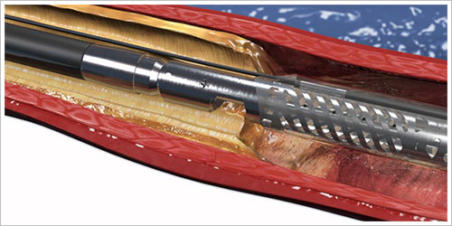
Rotablator
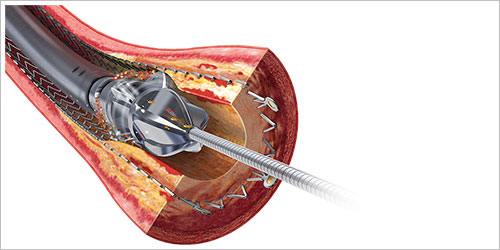
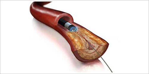
An angioplasty procedure is used to help relieve symptoms of angina. This is where a guide wire is passed into an artery in your heart that has become narrowed by a build- up of plaque.
A tiny balloon is inserted along the wire and then inflated to squash the plaque to the sides and improve the flow of blood through this section of the narrowed artery. This is often followed by the insertion of a metal mesh, called a stent, which holds the artery open after the balloon is withdrawn.
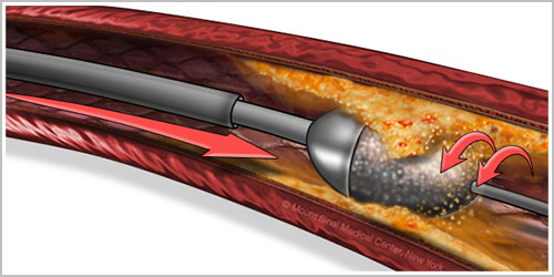
Sometimes, when the plaque is particularly hard, or is so narrow that the balloon can’t pass through it, rotablation may be used. Again, a very fine wire is guided through the narrowing.
After this, a special catheter (a thin tube) is inserted along the wire with a tiny drill at its tip, powered by compressed air. This drill is used to chip away at the plaque to gradually widen the narrowing. Once this has been done, a balloon can be inserted and the angioplasty can proceed as normal.
You’ll be awake for the procedure. Although the drill can be surprisingly noisy, it’s not painful, but as with an angioplasty, you may feel some minor chest discomfort. A local anaesthetic is used at the site in your groin or wrist where the catheter is inserted.
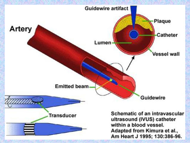
Intravascular Ultrasound (IVUS)
Intravascular Ultrasound (IVUS) is a catheter based system that allows physicians to acquire images of diseased vessels from inside the artery. IVUS provides detailed and accurate measurements of lumen and vessel size, plaque area and volume, and the location of key anatomical landmarks.